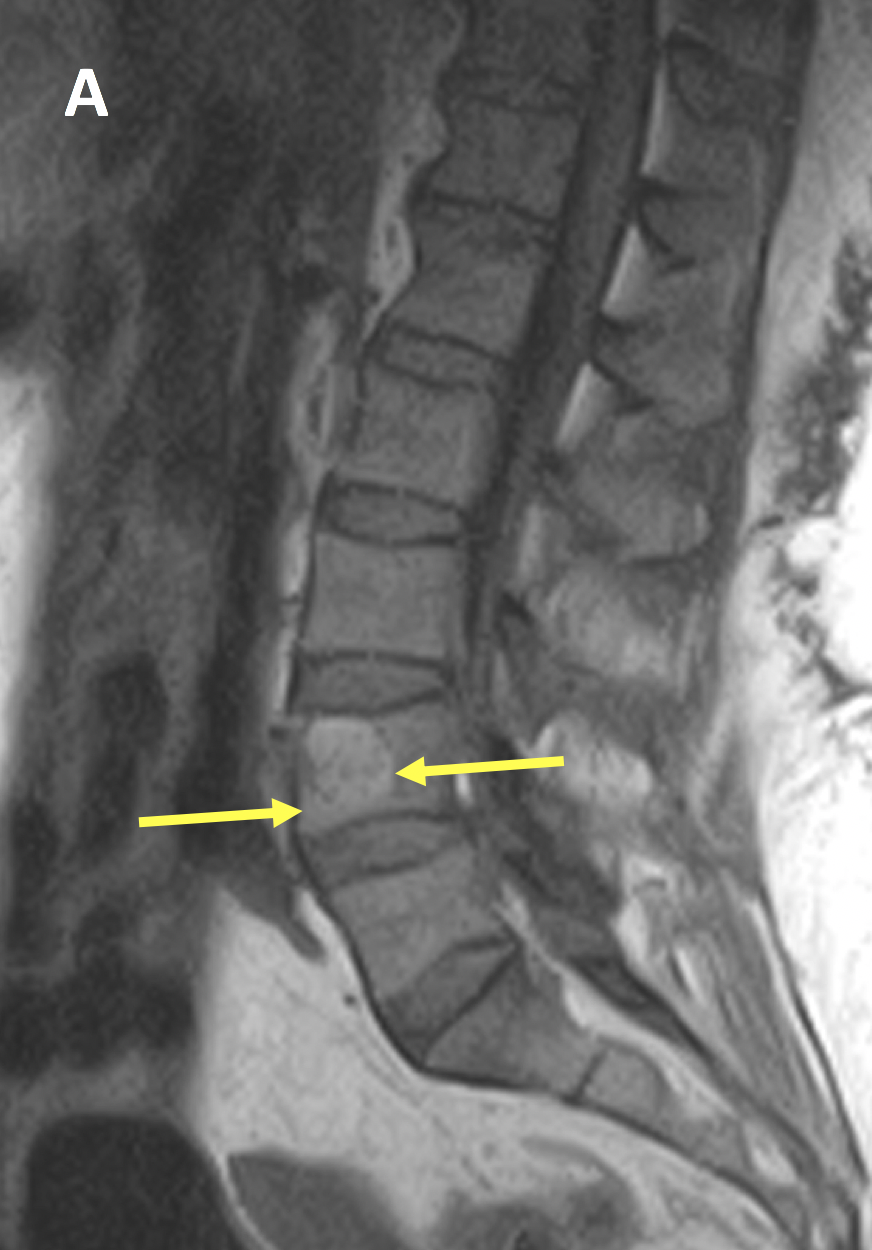
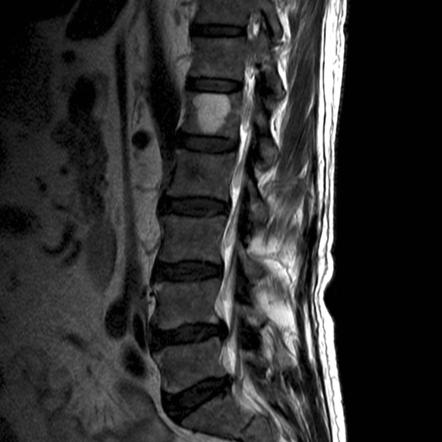
What Is A Benign Hemangioma Of The Spine
The typical location of the hemangioma in the spine. It is frequent enough in children.
 Vertebral Hemangioma Radsource
Hemangioma is a type of benign non-cancerous tumor formed due to an abnormal buildup of blood vessels.

What is a benign hemangioma of the spine. Spinal hemangioma also called vertebral hemangioma is a benign tumor that develops from blood vessels in the bones of the spine or vertebrae. They are benign in nature and frequently asymptomatic. Hemangioma of the spine is a benign tumor that affects the body of the vertebra.
What Symptoms Produce A Hemangioma. Your skin muscles internal organs or bones. A hemangioma of the spine is a benign tumor that is usually found in the mid-back thoracic and the lower back lumbar.
A hemangioma or haemangioma is a usually benign vascular tumor derived from blood vessel cell types. Theyre common and can occur anywhere in the body. The term hemangioma refers to a mass of blood vessels that commonly occur on the subcutaneous tissues.
Hemangiomas can also form on internal organs. 80 of the hemangiomas are cutaneous and consist of reddish raised patches on the skin. Spinal hemangiomas are benign tumors that are most commonly seen in the mid-back thoracic and lower back lumbar.
Given below is some information on what causes an atypical hemangioma in spine and how it can be treated. This HealthHearty write-up discusses the causes symptoms as well as the diagnostic methods and treatment options for this condition. These can originate pain in the area or they can be asymptomatic which means they do not produce any symptom.
Most cases show no symptoms. At The Spine Hospital at The Neurological Institute of New York we specialize in hemangiomas of the spine. Description Hemangiomas are benign bone lesions characterized by vascular spaces lined with endothelial cells.
The most common form is infantile hemangioma known colloquially as a strawberry mark most commonly seen on the skin at birth or in the first weeks of lifeA hemangioma can occur anywhere on the body but most commonly appears on the face scalp chest or back. A meningioma is a tumor that grows in the protective lining of the brain and spinal cord called the meninges. This type of tumor is most frequently diagnosed in patients between the ages of 30 and 50 and may not cause noticeable symptoms according to Scoliosis and Spine Associates.
Flawed vessels of the vertebra is formed inside the tumor. Most bone hemangiomas are on the spine and develop after age 50. Correct medical term is a hemangioma of the vertebra.
A vertebral hemangioma VH is a vascular lesion within a vertebral body. In fact they can be considered as benign tumors. The remaining lesions are found in the tibia femur and humerus.
A smaller proportion of patients with hemangioma manifested pain about 10. Pathological education is a benign tumor consisting of vascular tissue. Hemangiomas are benign tumors that develop from blood vessels.
A lumbar hemangioma is a benign blood vessel tumor that grows along one or more vertebra of the lower back. Hemangiomas most often appear in adults between the ages of 30 and 50. If they appear in other locations such as the spine liver complications can arise.
Signs of Thoracic Hemangioma The first step in treating a hemangioma is successful identification and diagnosis. Hemangiomas are noncancerous benign tumors made of abnormal blood vessels. They are very common and occur in approximately 10 percent of the worlds population.
Most are benign though in rare cases they can be cancerous malignant. A spinal hemangioma or a hemangioma in spine is a benign tumor that may develop in the bony segments of the spinal column. There are many types of hemangiomas and they can occur throughout the body including in skin muscle bone and internal organs.
While the tumor is not dangerous it can cause pain and discomfort and treatment may be recommended for these reasons. These growths classically appear in the thoracic and lumbar spine located in the mid to lower back. Vertebral hemangiomas are a common etiology estimated to be found in 10-12 of humans at autopsy.
Usually there is damage to the vertebral bodies but the possibility of tumor growth and cartilage layers. They occur in roughly 10 percent of the worlds adults and are considered quite common as a result. This disease can does not manifest itself and is often found only on surveys for other diseases.
A spinal hemangioma is a benign vascular tumor of the spine. Approximately 50 of osseous hemangiomas are found in the vertebral bodies thoracic especially and 20 are located in the calvarium. In the human body there are all in all 33 bones in the spine which are also known as vertebrae.
Most are benign and have no impact. Commonly these are benign lesions that are found incidentally during radiology studies for other indications. Thoracic hemangiomas are benign spinal tumors that most often impact adults aged 30 to 50.
Hemangioma is a vascular tumor which is a tangle of interwoven and modified vessels. A hemangioma is a benign noncancerous tumor made up of blood vessels. Hemangiomas of the skin are often visible as red raised birthmarks that generally fade on their own.
 Vertebral Hemangioma Radiology Case Radiopaedia Org
 Long Term Outcome Of Treatment Of Vertebral Body Hemangiomas With Direct Ethanol Injection And Short Segment Stabilization The Spine Journal
 Vertebral Hemangioma Radsource
 Vertebral Hemangioma Radsource
 Hemangioma Vertebral Mri Online
 Computed Tomography Of Thoracic Spine Showing T8 Vertebral Hemangioma Download Scientific Diagram
 Vertebral Hemangioma Radiology Reference Article Radiopaedia Org
 Metastatic Vertebral Lesion Mimicking An Atypical Hemangioma With Negative 18f Fdg Positron Emission Tomography Computed Tomography Sciencedirect
 Vertebral Hemangioma Radsource
Hemangioma In The Spine The Pain Source Makes Learning About Pain Painless
 Spine Lecture Hemangioma Of Spine Youtube
 Vertebral Hemangioma Wikipedia
 Benigni Benign Hemangioma Spine
 Metastatic Vertebral Lesion Mimicking An Atypical Hemangioma With Negative 18f Fdg Positron Emission Tomography Computed Tomography Sciencedirect
 Vertebral Hemangioma Radsource
 Atypical Mr Imaging Features Of Vertebral Hemangioma Involving The T12 Download Scientific Diagram
 Vertebral Hemangioma Radsource
 Lecture Vertebral Hemangioma Of The Spine